


BIOPHYSICS IS VERY COOL
A few weeks ago, we were asked to find ways on how Physics can help us understand Biology better. After much discussion, our group has finally found a way that can link Physics and Biology together. From there, we will be able to appreciate Biology with Physics.
We have come out with this link:
Concept of light (Physics) => How microscope works => Uses of microscope => Cells (Biology)
Concept of light
- defined as part of the electromagnetic spectrum that can be detected by the human eye
- travels in straight line at a speed of 3x10^8 m/s
- shadow formation(including solar eclipses) and the pin-hole camera illustrate the rectilinear propagation of light
- go through reflection and refraction
Introduction to microscope
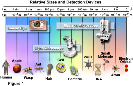
Ever since microscopes is invented in the late 1500s, light microscopes have enhanced our knowledge in medical areas and in the study of human and environmental science. Light microscopes can magnify objects up to more than 1,000 times, revealing microscopic details. Special techniques and optics have been developed to reveal the structures and biochemistry of living cells. The light microscope has greatly advanced our biomedical knowledge and continues to be a powerful tool for scientists.
Components of a typical microscope

Specimen control - hold and manipulate the specimen
- Stage - where the specimen rest
- Clips - used to hold the specimen still on the stage
- Micromanipulator - device that allows you to move the specimen in controlled, small increments along the horizontal direction
Illumination - shed light on the specimen (The simplest illumination system is a mirror that reflects room light up through the specimen.)
- Lamp - produces the light (Typically, lamps are tungsten-filament light bulbs. For specialized applications, mercury or xenon lamps may be used to produce ultraviolet light. Some microscopes even use laser to scan the specimen.)
- Rheostat - alters the current applied to the lamp to control the intensity of the light produced
- Condenser - lens system that aligns and focuses the light from the lamp onto the specimen
- Diaphragms or pinhole apertures - placed in the light path to alter the amount of light that reaches the condenser (for enhancing contrast in the image)
Lenses - form the image
- Objective lens - gathers light from the specimen
- Eyepiece - transmits and magnifies the image from the objective lens to your eye
- Nosepiece - rotating mount that holds many objective lenses
- Tube - holds the eyepiece at the proper distance from the objective lens and blocks out stray light
Focus- position the objective lens at the proper distance from the specimen
- Coarse-focus knob - used to bring the object into the focal plane of the objective lens
- Fine-focus knob - used to make fine adjustmets to focs the image
Support and alignment
- Arm - curved portion that holds all of the optical parts at a fixed distance and aligns them
- Base - supports the weight of all of the microscope parts
How microscope works in cooperation with the light?
The Basics
A microscope must gather light from a tiny area of a thin, well-illuminated specimen that is close-by. The objective lens of a microscope is small and spherical, which means that it has a much shorter focal length on either side. It brings the image of the object into focus at a short distance within the microscope's tube. The image is then magnified by a second lens, called an ocular lens or eyepiece, as it is brought to your eye.
How light travel through the microscope towards our eyes?
First of all, let us define refraction.
Refraction is the turning or bending of any wave, such as a light or sound wave, when it passes from one medium into another of different optical density. Refraction causes light to concentrate at one point thus focusing it.
The microscope contribute to the magnification of the specimens in fact, a light microscope has a white light source releasing rays that pass through the condenser lens to intensify the light and focus it on the sample. The specimen is placed on a slide, which is located on the stage of the microscope. The light travels through the specimen into the objective lens in which the specimen image is magnified. The light remains up through the body tube and reaches the ocular lens in which once again the image is magnified.
All the lens made up the microscope contribute to the magnification of the specimens.
The lenses magnify an object by bending the light that passes through them.
The ability to clearly distinguish individual parts of an object is resolution

Uses of microscope (Introduction of microscope to Biology)

Cells
All living organisms are made up of cells. A cell is the simplest units of living matter that can maintain life and reproduce themselves.
A human body is made up of numerous cells. When similar cells are grouped together, they will form tissue. An organ will then be formed when several different kinds of tissues are arranged together and perform a special function. Organs will then be arranged together and can perform complex functions for the body which are called system. Systems compose the human body.
Therefore, it is important to study the cells so as to understand the human body and other living matter.
There are two main groups of cells, Prokaryotic and Eukaryotic cells present in this world. They differ in their structure, reproduction and metabolism.
General information
Prokaryotic cells | Eukaryotic cells | ||
Examples | Bacteria | Plant cells | Animal cells |
Cellularity | Unicellular | Unicellular or Multi-cellular | |
Reproduction | Asexual- Binary fission | Asexual – mitosis & Sexual (depends on the species) | Sexual- Meiosis |
Shapes | Rod-shaped Round-shaped Spiral-shaped | Fixed shape – presence of cell wall | No fixed shapes |
Metabolism (Cellular respiration) | Oxygen (may be toxic or needed for metabolism), hydrogen, carbon dioxide, sulfur, sulfide (depends on the species) | Oxygen | |
Internal structure
Prokaryotic cells | Eukaryotic cells | ||
Examples | Bacteria | Plant cells | Animal cells |
Cell nucleus | X | √ | √ |
Cytoplasm | X | √ | √ |
Centrioles | X | X | √ |
Mitochondria | X | √ | √ |
Chloroplast | X | √ | X |
Cytoskeleton | √ | √ | √ |
Ribosomes | √ | √ | √ |
Vacuoles | Does not have any vacuole | One large central vacuole | Many small vacuoles |
Chromosomes | One long DNA strand | Many | Many |
Outer structure:
Prokaryotic cells | Eukaryotic cells | ||
Examples | Bacteria | Plant cells | Animal cells |
Cell wall | √ | √ | Χ |
Flagella | √ | Χ | √ |
Plasma Membrane | √ | √ | √ |

Terms used | Definitions |
Unicellular | Organisms composed of one cell only. |
Multi-cellular | Organisms composed of more than one cell. |
Asexual reproduction | A reproductive process that requires only one parent and results in an offspring that is identical to the parent. |
Binary fission | The form of asexual reproduction in single-celled organisms by which one cell divides into two cells of the same size. |
Mitosis | The process of cell division, which results in two cells with the same chromosome and DNA content as the original cell. |
Sexual reproduction | Production of offspring whose genetic constitution is a mixture of that of two potentially genetically different gametes. |
Meiosis | The process of cell division in which two nuclear divisions occur with only one chromosome replication. Each of the resulting gametes receives a haploid set of chromosomes. There are four haploid daughter cells resulting from this process. |
Centrioles | A pair of cylinder-like structures that are embedded in the centrosome (the microtubule-organizing centre in most animal cells). They replicate during interphase. As mitosis begins, the centrioles migrate to the two poles, where they organize microtubules for proper chromosome alignment and separation. |
Mitochondria | Found within the cytoplasm of eukaryotic cells. These organelles are responsible for the energy conversion of most of the cellular energy metabolites into adenosine triphosphate (ATP) by oxidative phosphorylation. |
Cytoskeleton | Made up of three kinds of protein filaments: -Actin filaments (8nm) -Intermediate filaments (10nm) -Microtubules (25nm) * Figures above are size in diameter. A network of fibers throughout the cell's cytoplasm that helps the cell maintain its shape and gives support to the cell. Cellular organelles are held in place by the cytoskeleton. |
Flagella | Long hair-like structures on a cell or microorganism enabling movement or manipulation. |
Examples of the cells in the world

Most familiar one would be the human cheek cells.
This is the 400x magnification of the cheek cells.

Bacillus bacteria at 1000x magnification

Elodia Leaf at 400x magnification. (Plant cell)
Conclusion
Most cells (animal and plant) range in size between 1 and 100 micrometers. They are only visible with the aid of a microscope. Therefore, scientists are able to use the microscope to see the different structure present in the cells and through that, they are able to analyze the different mutations happening in the cells. Thus, finding cure to those medical conditions such as, SARs and AIDs.
Credits:
Our information are taken from the different sites and edited by us.
Topics covered by:
- Light=Junjie
- How microscope works in cooperation with light?=Yanglin
- Uses of microscope = Donald
- Cells = Liling
If you are interested in the following topics, you can go to the following websites for more information.
Light
http://www.howstuffworks.com/light-microscope.htm/printable
http://www.southwestschools.org/jsfaculty/Microscopes/compoundscope.html
http://www.bookrags.com/research/microscope-wop/
http://www.bact.wisc.edu/microtextbook/index.php?module=Book&func=displayarticle&art_id=82
How microscope works in cooperation with light?
http://science.howstuffworks.com/light-microscope.htm
Uses of microscope (Introduction of microscope to Biology)
http://micro.magnet.fsu.edu/cells/index.html
Cells
http://www.iscid.org/encyclopedia/Types_of_Cells
http://www.cellsalive.com/toc_cellbio.htm
http://www.purchon.com/biology/cells.htm
http://biology.about.com/od/evolution/a/aa091004a.htm
http://micro.magnet.fsu.edu/cells/animalcell.html
http://www.britannica.com/eb/art-102103/Bacterial-cells-differ-from-animal-cells-and-plant-cells-in
http://www.danacode.co.il/thecel/cel1.htm
Labels: JLYD

posted by JLYD @ 10:20 PM
0 Comments
![]()